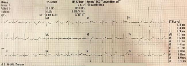
Normal Ecg Life In The Fast Lane

Assessment of the t wave represents a difficult but fundamental part of ecg interpretation.
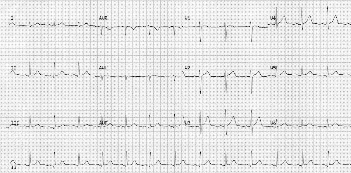
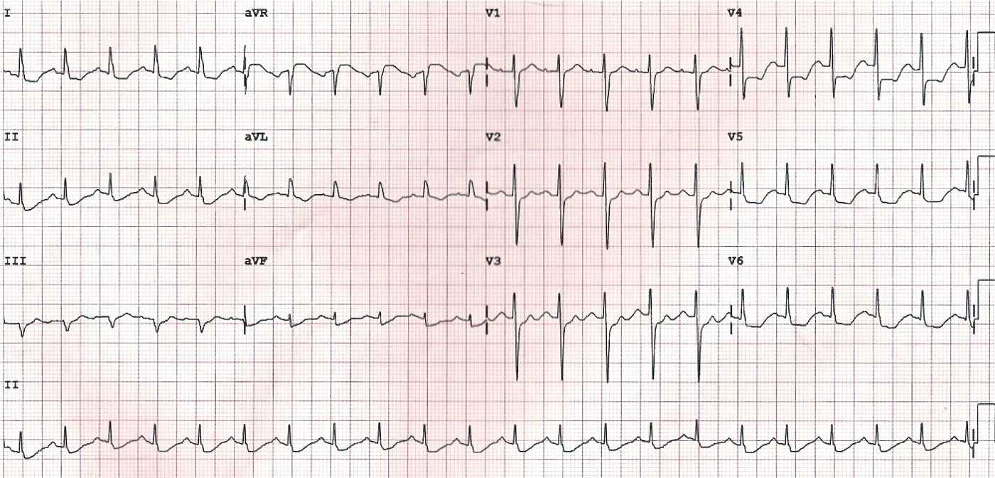
Normal ecg life in the fast lane. This supplies 10 of the ventricular filling at rest but up to 40 in tachycardia. Note that the heart is beating in a regular sinus rhythm between 60 100 beats per minute specifically 82 bpm. Normal p wave axis. Characteristics of normal sinus rhythm.
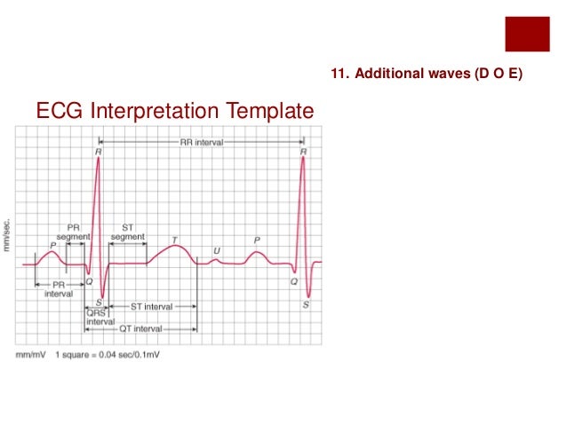
Normal qtc values qtc is prolonged if 440ms in men or 460ms in women qtc 500 is associated with increased risk of torsades de pointes qtc is abnormally short if 350ms. Luciani period 1873 luciani luigi. P waves should be upright in leads i and ii inverted in avr. Pr interval 120 ms suggests pre excitation the presence of an accessory pathway between the atria and ventricles or av nodal junctional rhythm.
Ecg mobitz av block mobitz 2nd degree av block atrioventricular block. In other words it s a normal finding with a secondary st t wave abnormality. The pr interval remains constant. The normal pr interval is between 120 200 ms 0 12 0 20s in duration three to five small squares.
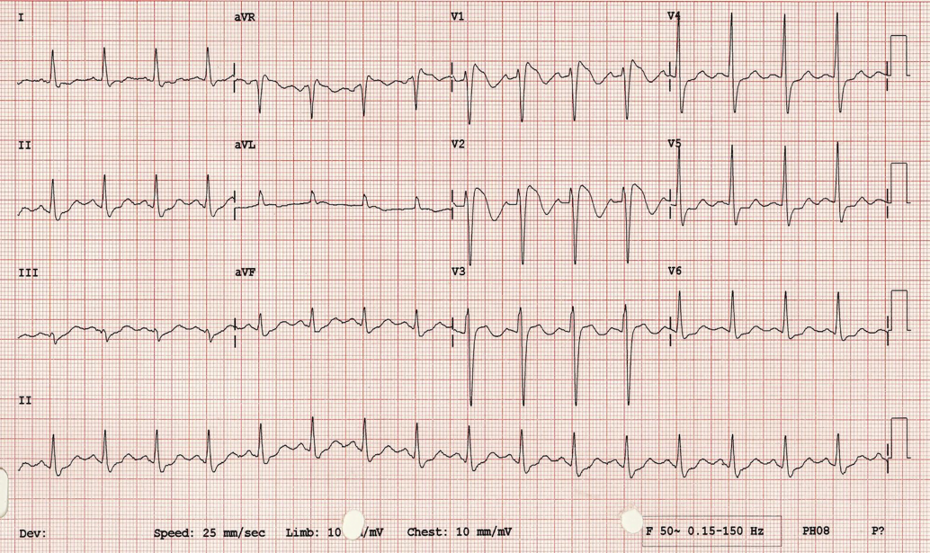
Regular rhythm at a rate of 60 100 bpm or age appropriate rate in children. The normal t wave in adults is positive in most precordial and limb leads. Generally speaking it means the t waves on the ecg are deflected opposite the majority of the qrs complex which is a normal finding in left bundle branch block paced rhythm and left ventricular hypertrophy with strain. Arterial pressure is still falling.
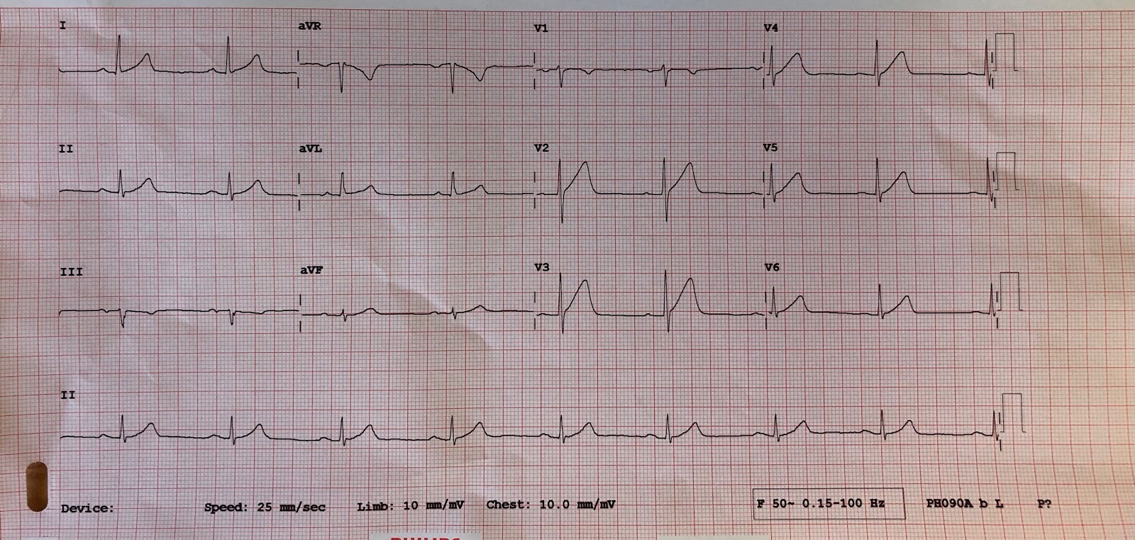
Since in the normal heart there is more myocardial mass to depolarize in the left ventricle than in the right the cardiac axis on ecg ends up pointing left between 0 and 90 degrees. A normal ecg is illustrated above. Each qrs complex is preceded by a normal p wave. Initial right axis on ecg is normal and resolves after the first 6 months of life an extreme superior axis axis of 90 to 180 degreesis seen with av canal or osmium primum atrial septal defects.
All the important intervals on this recording are within normal ranges. The atria contract and remaining blood in the atria is ejected into the ventricle. The amplitude diminishes with increasing age. The ecg will show the beginnings of a p wave at the end of this phase.
If the pr interval is 200 ms first degree heart block is said to be present. Upright in leads i avf and v3 v6. Normal duration of less than or equal to 0 11 seconds. As noted above the transition from the st segment to the t wave should be smooth.
Group beating second degree av block 2nd degree. First ecg in a human 1887 waller augustus desiré.
